
Immunosuppression Risk & Pathogen Profile Checker
Select Immune Defect Type
Select the primary immune branch being suppressed by medication or condition.
Primary Threats
Clinical Presentation
Atypical Symptoms
Diagnostic Gold Standard
Risk Summary & Management Strategy
Mortality/Risk Level
Prophylaxis Importance
CriticalSelect an Immune Defect Type
Choose a category from the left panel to view specific pathogen risks, atypical symptoms, and diagnostic requirements.
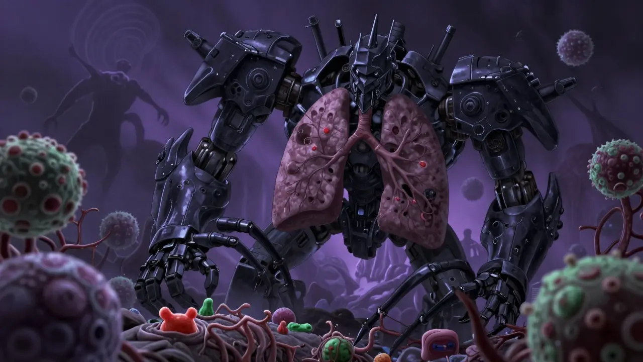
When your immune system is working correctly, it acts like a well-trained security team. It spots intruders, sounds the alarm, and kicks them out before they cause real trouble. But for patients on steroids or other immunosuppressants used to treat autoimmune diseases and prevent organ rejection, that security team is on vacation. This leaves the door open for organisms that most people never have to worry about. These aren't just common colds; we are talking about unusual, often aggressive pathogens that exploit specific gaps in your body's defenses.
The stakes are high because these infections don't always look the way you expect. A fever might be missing. The usual signs of inflammation could be absent. By the time symptoms appear, the infection may already be deep-seated and difficult to treat. Understanding which organisms target which immune defects is not just academic-it’s a matter of survival for anyone managing chronic immunosuppression.
Why Immunosuppression Changes the Rules
To understand the risk, you first have to understand what is being suppressed. Your immune system has different branches, each with a specific job. When medication dampens one branch, it creates a unique vulnerability profile. It’s not a blanket shutdown; it’s a targeted blind spot.
Humoral immunity (B-cells and antibodies) handles bacteria and some parasites. If this is weak, you become susceptible to things like Giardia intestinalis. On the other hand, Cell-mediated immunity (T-cells) manages viruses, fungi, and intracellular bacteria. Suppressing T-cells opens the floodgates for viruses like Cytomegalovirus (CMV) and fungi like Aspergillus. Research from the American Society of Transplantation highlights that patients with T-cell deficiencies face 15 to 20 times higher risk of viral reactivation compared to those with only B-cell issues.
Phagocyte defects, where the cells that eat bacteria don't work right, lead to severe skin and bone infections. You see high rates of Staphylococcus aureus and Gram-negative rods like Klebsiella and Pseudomonas in these cases. The key takeaway? Knowing your specific immune deficit helps predict your specific threats.
The Silent Threat: Atypical Presentations
In a healthy person, an infection screams for attention. You get hot, swollen, red, and painful areas. In an immunosuppressed patient, the infection whispers-or worse, stays silent. This is the most dangerous aspect of treating these patients.
A landmark study by Slatter et al. in 2007 looked at children awaiting hematopoietic stem cell transplantation (HSCT). They found that respiratory tract infections accounted for about 60% of all infections in this group. More startling was the finding that 23% of children who tested positive for pathogens via bronchoalveolar lavage (BAL) were completely asymptomatic. They had no cough, no fever, no shortness of breath. Yet, the bugs were there, multiplying.
This lack of classic inflammatory response means doctors cannot rely on traditional markers like fever or high white blood cell counts (leukocytosis) alone. Dr. Park’s 1967 case series in Archives of Dermatology showed early on how immunosuppressive therapy alters infection courses. One patient developed persistent varicella despite standard treatment, while another suffered severe herpes simplex with extensive tissue necrosis but lacked typical warning signs until it was too late. Another died from disseminated histoplasmosis that looked like simple skin inflammation rather than a systemic fungal invasion.
For patients, this means vigilance. You can’t wait for the obvious signs. Regular surveillance testing, especially for respiratory pathogens, becomes critical. If you feel slightly off, even without a fever, speak up. Early detection is the only advantage you have when your natural alarms are muted.
Unusual Respiratory Pathogens
The lungs are a primary battleground for immunosuppressed patients. While Pneumocystis jirovecii pneumonia (PCP) remains the most frequently isolated pathogen-accounting for 22% of positive samples in Slatter’s study-the list of potential invaders is long and varied.
Beyond PCP, you need to watch for:
- Viral Agents: Respiratory syncytial virus (RSV), influenza, parainfluenza, adenovirus, and human metapneumovirus. Even coronaviruses like NL63 and HKU1, which usually cause mild colds, can be severe here.
- Fungal Invaders: Aspergillus species are particularly deadly. According to the Infectious Diseases Society of America’s 2023 guidelines, Aspergillus infections in neutropenic patients have mortality rates exceeding 50%, even with optimal antifungal therapy. Compare that to 15% in immunocompetent patients, and the difference is stark.
- Parasitic Concerns: While less common in the lungs, systemic parasitic loads can impact respiratory function indirectly through overall debilitation.
The SARS-CoV-2 pandemic highlighted these vulnerabilities further. NIH case reports documented prolonged viral shedding exceeding 120 days in some immunocompromised patients, compared to the typical 10-14 days in healthy individuals. This prolonged presence increases the risk of mutation and transmission, making isolation protocols stricter for this group.
Gastrointestinal and Cutaneous Risks
Your gut and skin are also entry points for unusual organisms. For patients with humoral immunodeficiencies, such as X-linked agammaglobulinemia or selective IgA deficiency, Giardia intestinalis is a major concern. This flagellate protozoan causes chronic symptomatic infection in 87% of affected immunocompromised children, leading to foul-smelling stool, abdominal distension, and anorexia.
Treatment for Giardia is tricky. Metronidazole is the first-line drug (500 mg twice daily for 5-7 days), but failure rates jump to 30-40% in immunocompromised patients compared to 5-10% in healthy people. Often, combination therapy with tinidazole or nitazoxanide is required. Diagnosis requires more than a glance; stool microscopy combined with immunofluorescent antibody testing achieves 98% sensitivity, whereas standard methods might miss it.
Cutaneous infections manifest differently too. In phagocyte-deficient patients, you see granulomas forming at infection sites as T lymphocytes accumulate. About 73% of patients with chronic granulomatous disease develop hepatic or splenic microabscesses containing live intracellular organisms. Skin lesions may not heal, or they may spread rapidly, as seen in the historical cases of disseminated histoplasmosis presenting as widespread erysipelas-like inflammation.

Diagnostic Challenges and Strategies
Because symptoms are muted, diagnosis relies heavily on proactive testing rather than reactive observation. Waiting for culture results can cost precious time. Modern protocols emphasize rapid identification.
For respiratory issues, induced sputum has a sensitivity of only 65% for detecting PCP. Bronchoalveolar lavage (BAL), however, boosts that sensitivity to 92%. It’s more invasive, but for a patient whose life depends on catching an infection early, it’s worth it. For emerging threats, metagenomic next-generation sequencing (mNGS) is becoming a game-changer. It allows clinicians to identify pathogens in culture-negative infections, providing answers when traditional tests come back blank.
Prophylaxis plays a huge role. Without preventive measures, CMV infection rates can reach 40% in profoundly T-cell immunosuppressed allogeneic HSCT recipients. Preemptive therapy and targeted antimicrobials have helped, but infection-related mortality remains at 25-30% in this group according to the American Society of Hematology’s 2024 report. This underscores that prevention is not optional; it’s essential.
| Immune Defect Type | Primary Pathogens | Key Clinical Features | Diagnostic Gold Standard |
|---|---|---|---|
| Humoral (B-cell/Antibody) | Giardia intestinalis, Encapsulated Bacteria | Chronic diarrhea, foul stool, abdominal distension | Stool microscopy + Immunofluorescent antibody test |
| Cell-Mediated (T-cell) | CMV, Aspergillus, PCP, Adenovirus | Pneumonia, viral reactivation, tissue necrosis | Bronchoalveolar lavage (BAL), PCR |
| Phagocyte Defects | Staphylococcus aureus, Klebsiella, Pseudomonas | Skin/bone abscesses, granulomas, microabscesses | Culture, Biopsy |
| Severe Combined (SCID) | Mycobacterium avium, Mycobacterium bovis (BCG) | Disseminated mycobacterial disease, failure to thrive | Culture, Genetic Testing |
Emerging Therapies and Future Directions
The landscape is evolving. We are seeing promising developments in pathogen-specific T-cell therapies. Phase II trials show 70% response rates for refractory CMV and adenovirus infections, offering hope for patients who don’t respond to standard antivirals. Additionally, immunomodulatory approaches aim to temporarily enhance pathogen-specific immunity without triggering graft-versus-host disease in transplant recipients.
However, the core message remains unchanged. Immunosuppression is a double-edged sword. It saves lives by controlling autoimmune attacks or preventing organ rejection, but it introduces significant infection risks. Awareness, early testing, and strict adherence to prophylactic regimens are your best defenses against these unusual organisms.
What are the most common unusual organisms in immunosuppressed patients?
The most common unusual organisms include Pneumocystis jirovecii (causing PCP), Cytomegalovirus (CMV), Aspergillus species, and Giardia intestinalis. The specific organism depends on the type of immune defect; T-cell deficiencies favor viruses and fungi, while humoral defects favor certain bacteria and parasites like Giardia.
Why do immunosuppressed patients often lack fever during infections?
Fever is part of the inflammatory response mediated by cytokines and immune cells. Immunosuppressive drugs, such as steroids, blunt this inflammatory pathway. Consequently, patients may not mount a fever or show leukocytosis (high white blood cell count), even when severe infections are present, making clinical presentation atypical.
How does Aspergillus infection differ in immunosuppressed vs. healthy individuals?
In healthy individuals, Aspergillus often causes mild allergic reactions or self-limiting nodules. In immunosuppressed patients, particularly those who are neutropenic, it can cause invasive pulmonary aspergillosis with mortality rates exceeding 50%. The lack of immune containment allows the fungus to invade blood vessels and spread systemically.
What is the gold standard for diagnosing PCP in immunocompromised patients?
Bronchoalveolar lavage (BAL) is considered the gold standard for diagnosing Pneumocystis jirovecii pneumonia (PCP), with a sensitivity rate of approximately 92%. Induced sputum is less sensitive (around 65%) and may yield false negatives, delaying critical treatment.
Can Giardia be treated effectively in immunosuppressed patients?
Yes, but it is more challenging. First-line treatment is metronidazole, but failure rates are 30-40% in immunocompromised patients compared to 5-10% in healthy individuals. Combination therapy with tinidazole or nitazoxanide is often required to achieve clearance, and diagnosis should use high-sensitivity methods like immunofluorescent antibody testing.